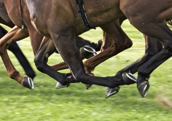
 Credit: Thinkstock Current technologies to assess fetlock health in horses have their limitations.
One cannot help but get excited about the possibilities for electroarthrography (EAG) as a diagnostic tool after speaking with Ontario Veterinary College researcher Mark Hurtig, DVM, MVSc, DACVS. He is developing a non-invasive way to assess joint cartilage health in fetlocks (the most commonly injured joint in horses).
Current technologies to assess fetlock health have their limitations. Veterinarians mainly use physical exams, diagnostic injections, X-ray images and ultrasound–yet these methods provide no information about the quantity or health of the articular cartilage that is critical for pain-free joint function.
Electroarthrography (EAG) is a novel method for easily assessing cartilage quality. Dime-sized electrodes are placed on the skin to record electrical signals produced by joint cartilage when loaded and unloaded. Researchers from the Université de Montréal and École Polytechnique reasoned that electrical signals might be measured on the skin surface similar to electrocardiography (ECG) for the heart. They found that people with knee arthritis had lower electrical potentials than normal people. So the concept of electroarthrography (EAG) was born. Hurtig’s contribution—with his team of postdoctoral fellows and graduate students—is applying the technique to horses by performing validation studies in cadaveric limbs, in which electrical signals can be directly correlated to cartilage quality.
Hurtig explained, “We thought that the fetlock might be a good place to start in the horse since the cartilage surface is close to the skin without any bulky muscles overlying the joint.”
Preliminary data from cadaveric forelimbs of horses under simulated weight bearing have shown that EAG signals can be easily recorded from the fetlock and are altered by damaged or osteoarthritic cartilage. When the cartilage is deliberately damaged with an enzyme like those found in osteoarthritic cartilage, it produces lower EAG signals. Once this validation study is complete, the next phase is to apply EAG to normal and lame horses.
In a preliminary live-animal test, electrical signals were recorded from fetlock joint cartilage while the horse was being pushed side to side while standing on a steel force platform. Human researchers have adopted the same technique using a Wii (game) platform. Software correlates the electrical signal on the skin surface of the knee to the timing of weight shift sensed by the platform. The strength of the EAG signal under the same weight indicates the status of cartilage health. In an eroded or damaged cartilage surface, the water and protein content changes resulting in a decline of electrical signals.
Since a horse is too heavy for Wii and a platform will not be portable for on-farm assessment, an instrumented horse boot (capable of recording weight bearing while the EAG signal is recorded) is being developed by other collaborators in the University of Guelph School of Engineering.
Future research will include localizing cartilage damage by loading different parts of the fetlock joint using wedge pads. Hurtig explaineds, “If we are successful it could mean that we can diagnose cartilage damage years before conventional methods.”
Electroarthrography could prove clinically useful in diagnosing hard to pinpoint lameness such as early osteoarthritis. Hurtig also pointed out that EAG could be used in similar contexts as diagnostic ultrasound in tendon injuries, which has been useful in establishing when it is safe to step up rehabilitation or return a horse to work.
This article was written by Jackie Bellamy-Zions. Funding for this research has been provided by Biomomentum Inc., Natural Sciences and Engineering Research Council of Canada (NSERC) and Equine Guelph.

