Ontario Veterinary College graduate student Cristin McCarty is no stranger to joint health issues, having to work through her own athletic injuries from rowing. As an avid rider since age 5, McCarty bought an ex-racehorse in her early teens for repurposing into a hunter/jumper and immediately began an education in joint problems and maintenance options aimed towards keeping her newly purchased horse sound.
McCarty’s interest in how osteoarthritis begins was born from experience. After pursuing an education in biology, McCarty was very excited to be given the opportunity to work with Dr. Jeff Thomason and Dr. Mark Hurtig at the University of Guelph, where ground-breaking research is looking at joint loading of horses traveling at high speed. Thomason specializes in biomechanics, studying mechanics of locomotion in horses, in-vivo bone strain and finite-element (FE) modeling of skeletal mechanics. Hurtig is an expert on the mechanical causes of osteoarthritis.
McCarty has been working with FE, gathering and analyzing data on loads acting at the fetlock joint and stresses in the cannon bone. By creating a computer-generated model using computed tomographic (CT) or MRI images of an equine fetlock joint they are working on determining the internal bone stress under varying loading conditions (rates, directions and magnitudes) using FE software. This method of analysis could provide further insight into the biomechanical role impact has on the stress distribution in areas of high remodeling, which are associated with osteoarthritis in racehorses.
FE has been used for quite some time in the automotive industry in crash test simulations to assess material failure but it is a relatively new technology for applications in biology. McCarty, who has been working under Thomason for three years now, said, “It was a steep learning curve to become familiar with the software to build complex models. That alone took almost two years.”
FE, through complicated mathematical calculations, can test where stress points will occur under particular loading conditions. In a more dense bone (which occurs in osteoarthritis), these calculations can show how stress points may be transferred and put onto the cartilage of the joint. Using FE researchers can also study how distribution of stress in a healthy, spongy less dense bone differs from that of an osteoarthritic joint.
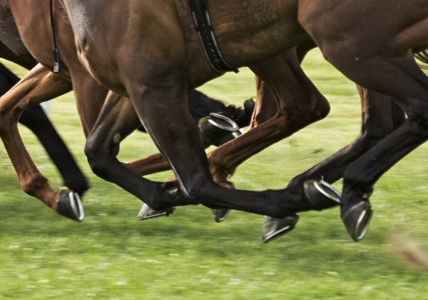
Contributing factors to osteoarthritis (OA) in the fetlock joint of a horse lies partly in conformation. A horses’ large body mass is held up by four fairly small limbs and the fetlock joint is a small area to distribute the force and loading that occurs during high speed movement. Long pasterns and steep joint angles increase stresses on the back of the leg and can predispose a horse to issues. McCarty explains the incredible forces calculated from the horses mass x acceleration (F=M x A) during the high speed work a racehorse performs. This can result in upwards of 2.5 times their body weight on one limb. Footing plays a role with harder surfaces resulting in higher strains on the hoof. A blunt force trauma could also predispose a horse to joint injury and make it more susceptible to the onset of OA.
Intense continuous training can set a horse up for OA and joint disease. McCarty goes on to talk about training programs and compares the training of human athletics to how we train race horses: “If you wanted to run a 200 meter sprint, you would not sit on a couch all day; get up to sprint 50 meters then return directly to the couch. With no pre-conditioning of joints, this is asking for injury. So why would we ask a horse to stand in a stall all day and then take them out for a gallop?” Chronic overloading of joints leads to problems over time.
One of the potential uses McCarty cites for FE modeling could be assessing the chronically lame horse. This technology may allow predications of where the bone may fracture and under what conditions. Also, with greater understanding of high stress point areas and what is creating them, mitigation may be possible such as suggesting a change in the footing surface the horse trains on.
This research will be continuing in collaboration with the Robarts Research Institute, Western University and has been funded by the Ontario Veterinary College Department of Biomedical Science, Ontario Ministry of Agriculture and Food, Natural Sciences and Engineering Research Council of Canada (NSERC).
Promote Healthy Joints
- Daily pasture turn out allows horses to load joints as nature intended
- Gradual, progressive warm up and cool down periods in training sessions
- Avoid excessive fast work and training on hard ground
- Exposing horses to different surfaces while training and hill work can be beneficial
- Contact your vet if you see swelling, lameness, shortened stride or any signs of pain
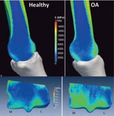
Healthy and Osteoarthritic Bone Density (Green = higher density bone). Mapped material stiffness on third metacarpal from micro-computed tomography images. Bottom images show the internal stiffness distribution through the medial/lateral plane of the distal end of MC3.
Learn more about osteoarthritis by visiting Equine Guelph’s healthcare tool “Journey through the Joints” and test your ability to spot lameness with “Lameness Lab.” Equine Guelph thanks Zoetis for sponsoring these valuable tools. For further information, visit www.EquineGuelph.ca.

