What do you do when your horse develops a lameness?If the lameness is pronounced or persistent, you will call your equine veterinarian.Your veterinarian is an expert in movement and anatomy, but he or she may recommend diagnostic imaging to better understand the nature and extent of an injury.More information correlates to more effective treatments and shorter recovery times for your horse.
In this article we will review the most common diagnostic imaging tools that are available in the equine veterinary market.Some of these tools, such as digital x-ray and ultrasound, are common in most veterinary practices.Other technologies such as fluoroscopy, scintigraphy and MRI are only available at some referral and specialty equine practices.
Digital x-rayDigital x-ray is a common imaging technology that can be used in the hospital or in the field.A quality x-ray image can detect structural problems such as broken bones, some soft tissue injuries, and foreign bodies such as nails in the sole.Generally, x-ray is not a costly procedure.
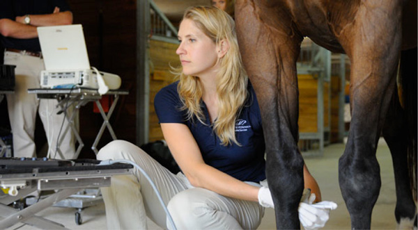
UltrasoundUltrasound technology uses non-audible sound waves to bounce off tissues to create images.Ultrasonography is better than x-ray at imaging soft tissue problems such as swelling and injuries inside a joint.
While many veterinarians offer x-ray and ultrasound, a few offer more specialized imaging to find damage that cannot be observed with the usual tools.
Nuclear ScintigraphyThis fascinating technology involves injecting a radioisotope dye into your horse’s bloodstream.The dye is then selectively concentrated into tissues that are inflamed.These inflamed areas show up on an image, allowing your veterinarian to diagnose stress and prevent further injury.Scintigraphy is an expensive procedure that requires haul in, sedation, and keeping your horse until he has metabolized the dye.
FluoroscopyFluoroscopy is a tool that creates live, real-time, video images of the structure inside your horse’s injured leg.The veterinarian can rotate and adjust the machine as necessary for the best view.
Magnetic Resonance Imaging (MRI)Occasionally, there is a need for an imaging tool that can do what no other tool can.MRI is such a tool.MRIs produce detailed, three-dimensional images of complex body structures.They are especially good for viewing the structures within a horse’s hoof.While there are several types of MRI equipment, not many practices offer MRI.The two most common types of MRI equipment include standing MRI, which is designed to allow the horse to remain upright during the imaging procedure, and mobile MRI, which is performed inside a trailer.This procedure requires full anesthesia of the horse.
While we hope that your horse has a long and healthy life on four sound legs, it is comforting to know that today’s imaging tools allow for more accurate diagnoses.Take the time to learn about the technologies that your veterinarian offers.It is important to know your options, so you can get back in the saddle.
Heather E. Lewis joined Animal Arts in August 2000 and has been a principal in the firm since 2004. Her primary area of expertise is the design and management of equine and large animal projects. Heather speaks regularly about the design of large animal facilities at the Veterinary Economics Hospital Design Conference for the Central Veterinary Conference. Heather spoke at the 2012 American Association of Equine Practitioners conference on “the equine hospital of the future.” She has been published on large animal facility design topics in Blackwell’s Five-Minute Veterinary Practice Management Consult and Veterinary Practice News.

